


 English
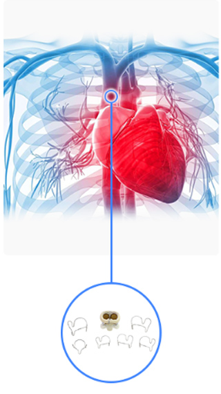
Embolization Coils are a cornerstone in interventional radiology and minimally invasive surgical procedures. They are small, specialized medical devices designed to treat abnormal blood flow, stop hemorrhages, or block vessels feeding tumors and aneurysms. Understanding how Embolization Coils work involves exploring their design, types, deployment techniques, physiological mechanisms, and clinical applications. In this article, we provide a detailed explanation suitable for medical professionals, students, and anyone interested in modern medical devices.
What Are Embolization Coils?
An embolization coil is a tiny, flexible wire—usually made of platinum, stainless steel, or other biocompatible metals—that is introduced into blood vessels to obstruct blood flow. These coils are inserted via a catheter under imaging guidance, often in procedures called transcatheter embolization.
Embolization coils serve multiple purposes:
Treating Aneurysms: Prevent rupture by filling the aneurysm sac.
Controlling Bleeding: Stop hemorrhages in trauma or gastrointestinal bleeding.
Tumor Treatment: Block blood supply to tumors in procedures like transarterial chemoembolization (TACE).
Vascular Malformations: Close abnormal vascular connections such as arteriovenous malformations (AVMs).
The principle behind embolization coils is to induce hemostasis or vascular occlusion by promoting clot formation, physically blocking the vessel, and redirecting blood flow.
Structure and Materials of Embolization Coils
Embolization coils vary in design depending on their intended clinical use. Key components include:
1. Core Material
Platinum: Highly radiopaque, allowing visualization under fluoroscopy.
Stainless Steel: Strong and flexible but slightly less radiopaque than platinum.
Nickel-Titanium (Nitinol): Provides shape memory and superelasticity for precise deployment.
2. Coil Shape
Helical or Spiral Coils: Traditional design for general vessel occlusion.
Complex 3D Coils: Pre-shaped to conform to aneurysms and prevent coil migration.
Detachable Coils: Can be repositioned or retrieved before final deployment.
3. Fibers or Attachments
Some coils are coated with synthetic fibers (e.g., Dacron, polyester) or hydrogel to accelerate clot formation and improve occlusion efficiency.
How Embolization Coils Work: Mechanisms
The effectiveness of embolization coils relies on a combination of mechanical blockage and biological response.
1. Mechanical Occlusion
Once deployed, the coil physically occupies space within the vessel or aneurysm sac, reducing blood flow. The coil’s shape and flexibility allow it to conform to the vessel walls, preventing migration and maximizing contact.
2. Thrombogenicity
Many coils are designed to enhance clot formation. Fibers or hydrogel coatings promote platelet adhesion and fibrin deposition, accelerating thrombus formation around the coil. This thrombotic occlusion ensures the vessel remains closed and reduces the risk of bleeding recurrence.
3. Remodeling and Healing
Over time, the occluded vessel undergoes fibrotic remodeling. The body gradually replaces the clot with fibrous tissue, permanently sealing the vessel. In aneurysm treatment, this prevents further expansion or rupture.
Types of Embolization Coils
Embolization coils are classified based on material, shape, and deployment mechanism:
1. Pushable Coils
Simple, non-detachable coils pushed through the catheter using a wire pusher.
Once released, they cannot be repositioned.
Suitable for vessels with straightforward anatomy.
2. Detachable Coils
Can be repositioned or retrieved before final deployment.
Offer precise control for aneurysms and complex vascular anatomy.
Deployment may involve mechanical, electrical, or electrolytic detachment systems.
3. Fibered vs. Non-Fibered Coils
Fibered coils: Promote faster thrombosis due to added fibers or coatings.
Non-fibered coils: Rely mainly on mechanical occlusion; used when slower clot formation is sufficient.
4. Hydrogel-Coated Coils
Swell upon contact with blood, enhancing occlusion.
Often used for aneurysms or large vessels requiring high filling density.
Deployment of Embolization Coils
Deploying embolization coils is a minimally invasive procedure performed by interventional radiologists or endovascular surgeons.
Step 1: Vascular Access
A catheter is inserted through an access site, often the femoral or radial artery.
Fluoroscopy guides the catheter to the target vessel.
Step 2: Coil Selection
The appropriate coil size and type are chosen based on vessel diameter and length.
Oversizing slightly helps ensure secure placement and reduces the risk of migration.
Step 3: Coil Introduction
The coil is advanced through the catheter to the target site.
For detachable coils, deployment is paused if repositioning is needed.
Step 4: Coil Placement and Occlusion
The coil is released into the vessel, conforming to the vessel walls or aneurysm sac.
Blood flow slows, and thrombus formation begins.
Additional coils may be deployed to achieve complete occlusion.
Step 5: Verification
Angiography is performed to confirm vessel closure and assess blood flow redistribution.
The catheter is then removed, and the access site is closed.
Physiological Response to Embolization Coils
Once in place, embolization coils trigger a series of biological events:
Immediate Mechanical Effect: Blood flow is reduced by the coil’s physical presence.
Thrombus Formation: Platelets and fibrin accumulate around the coil.
Inflammatory Response: Local tissue responds to the coil and thrombus formation.
Fibrotic Remodeling: The thrombus is replaced with permanent fibrous tissue, securing vessel closure.
This combination of mechanical and biological effects ensures long-term efficacy of embolization therapy.
Clinical Applications
1. Cerebral Aneurysms
Coils are deployed into aneurysm sacs in the brain to prevent rupture.
Detachable coils are preferred due to the need for precise placement.
2. Gastrointestinal Bleeding
Embolization coils can stop bleeding in arteries supplying the stomach, intestines, or liver.
Minimally invasive alternative to open surgery.
3. Trauma
Coils quickly occlude injured vessels in trauma patients, controlling hemorrhage and stabilizing the patient.
4. Tumor Treatment
Transarterial chemoembolization (TACE) uses coils to block blood supply to liver tumors, allowing targeted chemotherapy and tumor necrosis.
5. Arteriovenous Malformations
Coils occlude abnormal connections between arteries and veins, preventing hemorrhage and correcting blood flow patterns.
Advantages of Embolization Coils
Minimally Invasive: Reduced recovery time compared to open surgery.
Precision: Detachable coils allow accurate placement.
Effectiveness: High success rate in aneurysm and hemorrhage control.
Versatility: Used in various vascular conditions, from trauma to oncology.
Long-Term Durability: Induces permanent vessel closure through thrombus formation and fibrosis.
Potential Risks and Complications
Although embolization coils are generally safe, some risks include:
Coil migration or misplacement
Vessel perforation
Non-target embolization leading to tissue ischemia
Allergic reactions to coil material (rare)
Recanalization or incomplete occlusion in some cases
These risks are minimized by careful imaging guidance, proper coil selection, and experienced operators.
Innovations in Embolization Coil Technology
Recent advancements have improved coil performance and safety:
Bioactive Coils: Coated with materials promoting faster endothelialization.
Hydrogel-Coated Coils: Swell in situ to increase occlusion efficiency.
Shape-Memory Coils: Nitinol-based coils that conform to complex aneurysm geometry.
Detachable Electrolytic Coils: Precise deployment with controlled detachment for delicate vascular structures.
These innovations expand the range of clinical applications and improve patient outcomes.
Conclusion
Embolization coils are powerful medical tools that work by combining mechanical blockage and biological thrombosis to occlude blood vessels safely and effectively. From treating cerebral aneurysms to controlling gastrointestinal bleeding and supporting tumor therapy, how embolization coils work is a fascinating interplay of engineering, material science, and human physiology.
Advancements in coil materials, design, and deployment techniques continue to enhance the precision, safety, and versatility of embolization procedures. For clinicians, understanding the mechanisms, proper use, and potential complications is crucial for achieving optimal patient outcomes.
As minimally invasive procedures become increasingly preferred, embolization coils remain a critical component of modern interventional radiology, demonstrating the profound impact of small devices on life-saving treatments.
